

Software for CADEYE


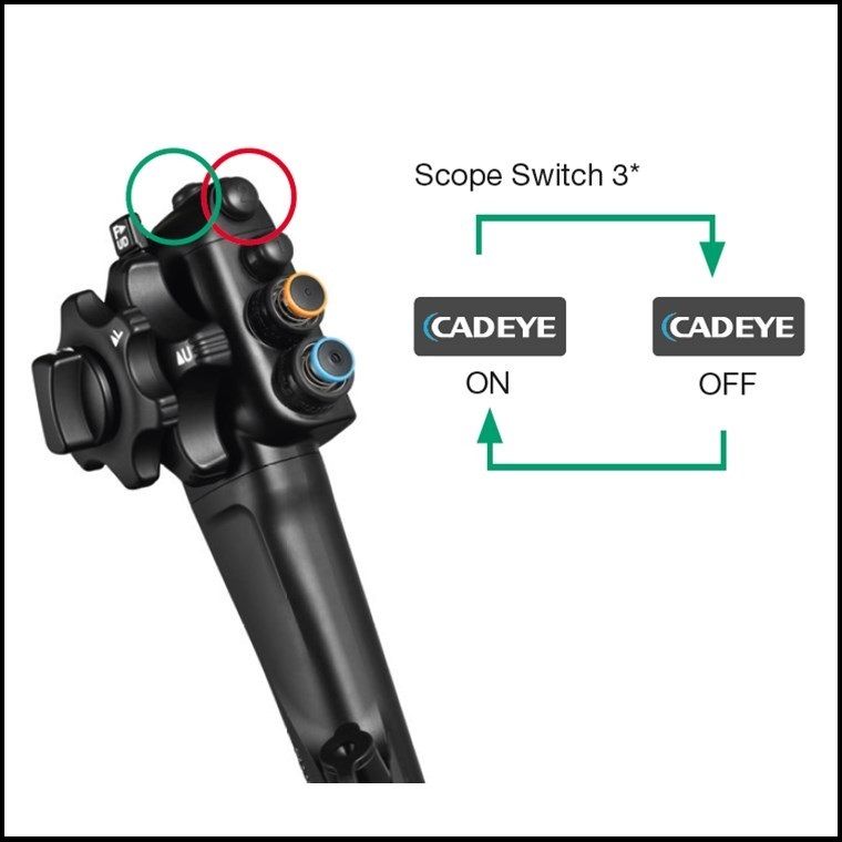
Seamless Operation
CAD EYE Detection can be activated / deactivated simply by the push on the endoscope button or directly at the processor.
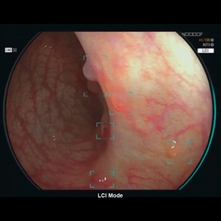
CAD EYE Detection Box
CAD EYE was developed to support real time detection of colonic polyps utilising AI technology. When a suspicious polyp is detected within the endoscopic image, a Detection Box indicates the area where the suspicious polyp has been detected accompanied by a sound signal.
Characterisation Support
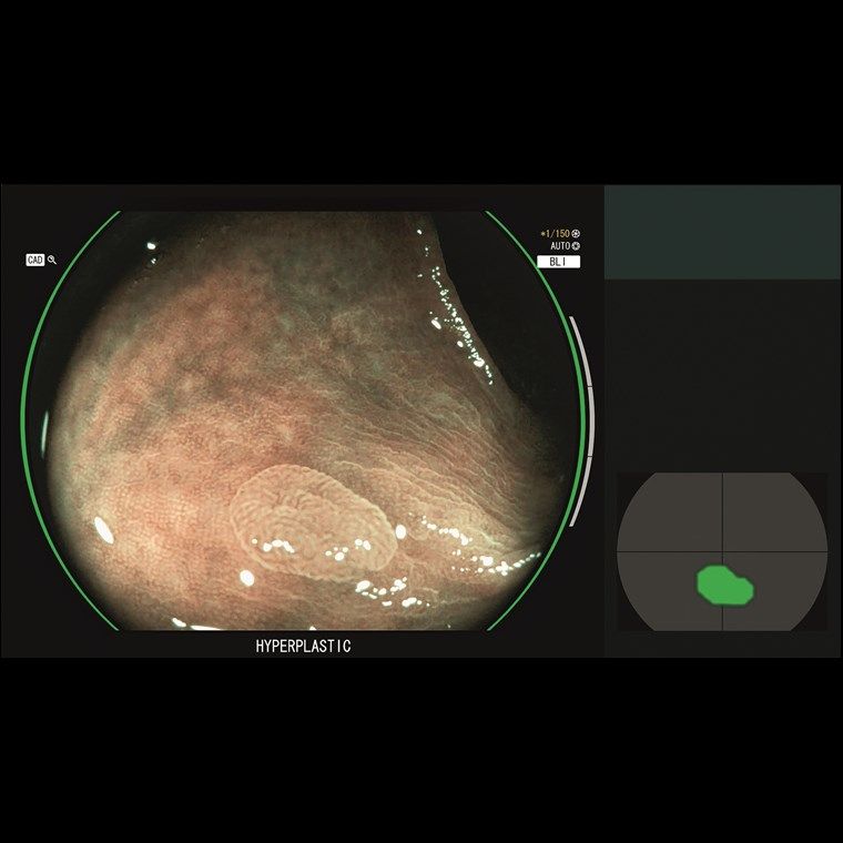
Once a suspected polyp is detected by CAD EYE Detection (WLI or LCI), CAD EYE Characterisation - in combination with BLI - can support endoscopists in the diagnosis of the polyp. This function analyses in real-time and without freezing or zooming if a polyp is hyperplastic or neoplastic, which is visually indicated by the use of different colour codes in the Position Map.
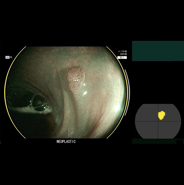
CAD EYE Detection with LCI
CAD EYE Characterisation is aimed to make procedures more efficient by increasing the accuracy of diagnosis to expert-level. GREEN: Characterisation HYPERPLASTIC; YELLOW: Characterisation NEOPLASTIC.
CAD EYE works with the expansion unit EX-1 and the CAD EYE software EW10-EC02 and can store up to 30 hours of video material in its internal memory. It can easily be controlled with the scope switch or directly at the processor.
| EX-1 Expansion Unit | |
|---|---|
| Compatible Processors | VP-7000 / EP-6000 |
| Compatible Scopes | 700 Series colonoscopes |
| Dimensions (W x H x D) | 370.0 x 99.0 x 456.6mm |
| Weight | 7.1 kg |
| Output | DVI-I x1, DVI-D x1 |
| Input | DVI-I x1 |
| Memory | 30 hours of video material, Full-HD, MP4 |
| Software EW10-EC02 | |
|---|---|
| Package Content | USB flash drive for CADEYE installment, user manual |
Brand: FujiFilm |
Code: EW10EC02V1
MPN:
APN: 9319499195502 |
Supplier Code:

Brand: FujiFilm |
Code: EW10EC02V1
MPN:
APN: 9319499195502 |
Supplier Code:
At a glance
CAD EYE‘s functionality is available with the compatible expansion unit EX-1, including software EW10-EC02 in combination with the Fujifilm ELUXEO 7000 system and 700 series colonoscopes. The internal memory allows the storage of up to 30 hours video material.
Seamless OperationCAD EYE Detection can be activated / deactivated simply by the push on the endoscope button or directly at the processor. |
|
|
|
CAD EYE Detection BoxCAD EYE was developed to support real time detection of colonic polyps utilising AI technology. When a suspicious polyp is detected within the endoscopic image, a Detection Box indicates the area where the suspicious polyp has been detected accompanied by a sound signal. |
Characterisation SupportOnce a suspected polyp is detected by CAD EYE Detection (WLI or LCI), CAD EYE Characterisation - in combination with BLI - can support endoscopists in the diagnosis of the polyp. This function analyses in real-time and without freezing or zooming if a polyp is hyperplastic or neoplastic, which is visually indicated by the use of different colour codes in the Position Map.
|
|
|
|
CAD EYE Detection with LCICAD EYE Characterisation is aimed to make procedures more efficient by increasing the accuracy of diagnosis to expert-level. GREEN: Characterisation HYPERPLASTIC; YELLOW: Characterisation NEOPLASTIC. |
FUJIFILM has launched a new software version with a new function for colon polyp characterisation utilising a type of Artificial Intelligence (AI) called deep learning. FUJIFILM Corporation had already obtained a CE mark for a previous software version with the colon polyp detection function in February 2020, and both the detection and the new characterisation function have been named CAD EYE™.
The new characterisation functionality of CAD EYE together with the polyp detection function is available with software EW10-EC02 and the compatible expansion unit EX-1 in combination with Fujifilm’s ELUXEO 7000 system and the 700 series colonoscopes. Fujifilm has already obtained a CE mark for EW10-EC02.
The new CAD EYE Characterisation will assist clinicians by generating a suggested histological prediction by displaying whether the suspicious polyp(s) in the image are hyperplastic or neoplastic.
CAD EYE functions while the moving endoscopy image is being observed and does not require complicated techniques or operations such as magnification and image capturing.
Prior to CAD EYE, Fujifilm had developed two different types of image enhancement technologies called LCI (Linked Colour Imaging) and BLI (Blue Light Imaging) for supporting detection and characterisation respectively due to their characteristics of light wavelength used. Fujifilm has applied this idea to the development of CAD EYE, and CAD EYE’s functionalities are automatically activated depending on the current observation mode.
CAD EYE Detection is activated when the clinicians are observing in White Light Mode or LCI Mode, and it automatically switches to CAD EYE Characterisation when the observation mode is changed to BLI Mode. CAD EYE can be activated or deactivated simply with just one click of the scope switch, which is also considered important when the functions are no longer necessary such as during therapeutic procedures.

