Artificial Intelligence for Fujifilm ELUXEO System


Real Time Detection
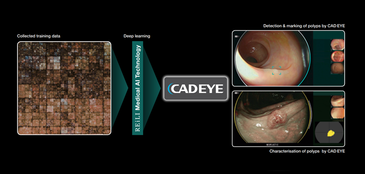
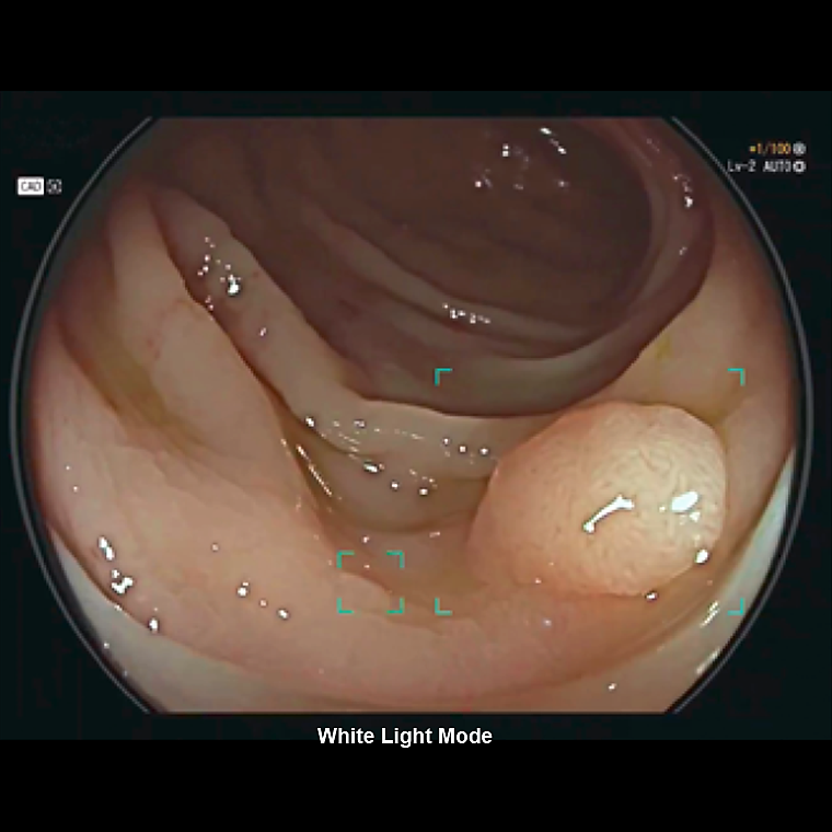
CAD EYE is aimed to improve the real time polyp detection rate to expert level, helping to recognise flat lesions, multiple polyps simultaneously as well as any lesions at the corner of the image. CAD EYE Detection is possible with White Light and LCI (Linked Color Imaging) mode.
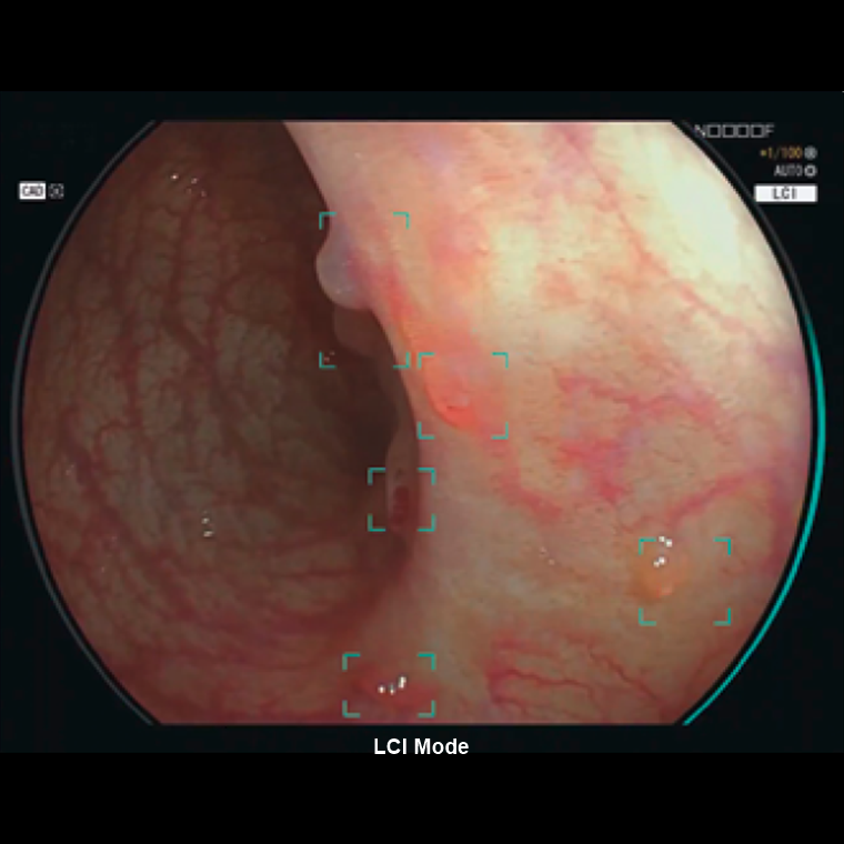
CAD EYE Detection with LCI
CAD EYE for colonic polyp detection is aimed to improve the adenoma detection rate to expert level.
Characterisation Support
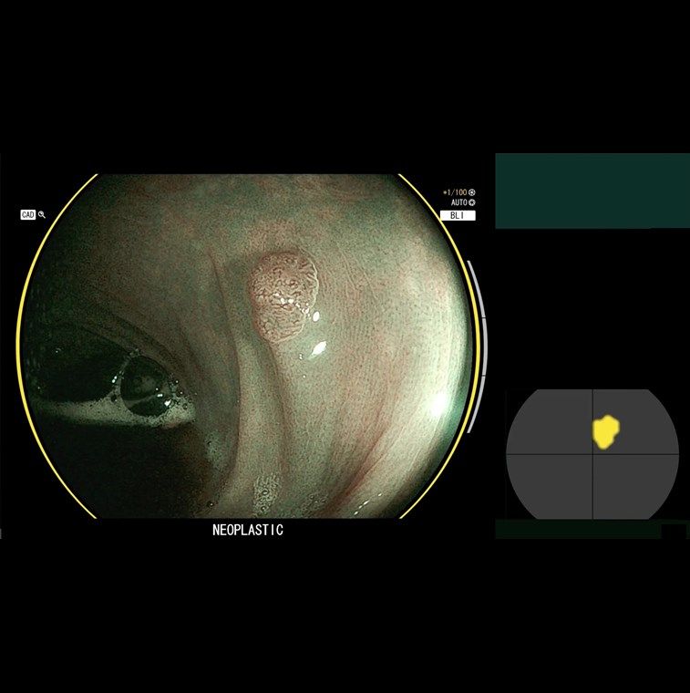
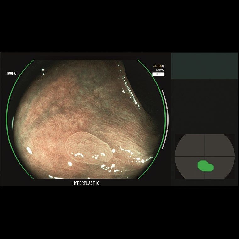
Once a suspected polyp is detected by CAD EYE Detection (WLI or LCI), CAD EYE Characterisation - in combination with BLI - can support endoscopists in the diagnosis of the polyp. This function analyses in real-time and without freezing or zooming if a polyp is hyperplastic or neoplastic, which is visually indicated by the use of different colour codes in the Position Map.
CAD EYE Detection with LCI
CAD EYE Characterisation is aimed to make procedures more efficient by increasing the accuracy of diagnosis to expert-level. GREEN: Characterisation HYPERPLASTIC; YELLOW: Characterisation NEOPLASTIC.
User Interface
The development of the user-friendly interface has been pursued to enable comfortable procedures. It does not interfere with clinical images and minimises required eye movement. Its display is designed to be simple and intuitive for excellent support during long hours in the examination room.
Interface Menu
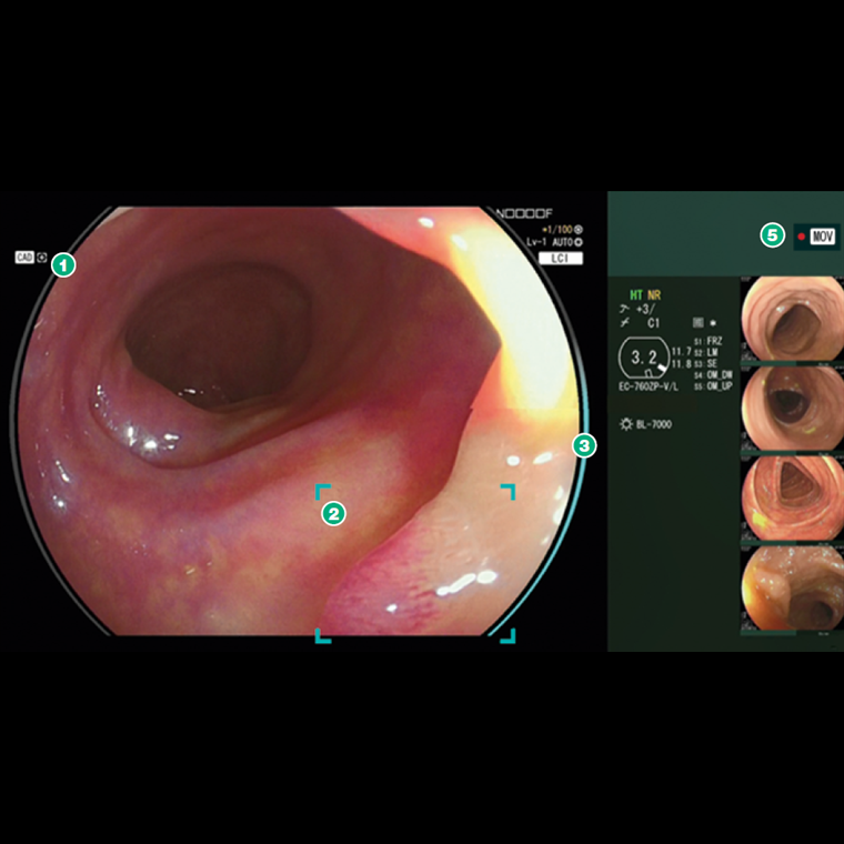
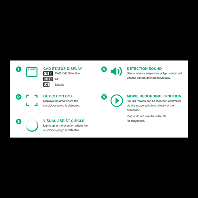
- 1. CAD Status Display - Indicates status of characterisation analysis regarding to area suspected.
- 2. Detection Box - Displays the area where the suspicious polyp is detected.
- 3. Visual Assist Circle - Lights up in the direction where the suspicious polyp is detected.
- 4. Detection Sound - Sound signal when a suspicious polyp is detected. Volume can be defined for each user.
- 5. Movie Recording Function - Full HD movies can be recorded, controlled by the scope switch or directly at the processor.
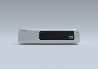
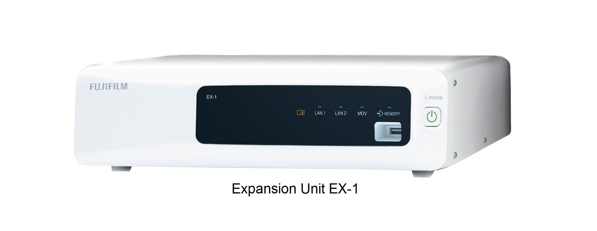
CAD EYE works with the expansion unit EX-1 and the CAD EYE software EW10-EC02 and can store up to 30 hours of video material in its internal memory. It can easily be controlled with the scope switch or directly at the processor.
| EX-1 Expansion Unit | |
|---|---|
| Compatible Processors | VP-7000 / EP-6000 |
| Compatible Scopes | 700 Series colonoscopes |
| Dimensions (W x H x D) | 370.0 x 99.0 x 456.6mm |
| Weight | 7.1 kg |
| Output | DVI-I x1, DVI-D x1 |
| Input | DVI-I x1 |
| Memory | 30 hours of video material, Full-HD, MP4 |
| Software EW10-EC02 | |
|---|---|
| Package Content | USB flash drive for CADEYE installment, user manual |
| Brochures | |
|---|---|
| ELUXEO CADEYE - Brochure | Datasheets |
| Artificial Intelligence for Fujifilm ELUXEO System - Datasheet |
Brand: FujiFilm |
Code: CADEYE
Supplier Code: EX-1 + EW10-ECO2 V1
Supplier Description: Expansion Unit incl. SW

Brand: FujiFilm |
Code: CADEYE
Supplier Code: EX-1 + EW10-ECO2 V1
Supplier Description: Expansion Unit incl. SW
At a glance
CAD EYE has been developed utilising AI deep learning technology and is compatible with Fujifilm’s ELUXEO™ endoscopy series to support endoscopic lesion detection and characterisation in the colon. Kit Contents
Real Time DetectionCAD EYE is aimed to improve the real time polyp detection rate to expert level, helping to recognise flat lesions, multiple polyps simultaneously as well as any lesions at the corner of the image. CAD EYE Detection is possible with White Light and LCI (Linked Color Imaging) mode. |
|
|
|
CAD EYE Detection with LCICAD EYE for colonic polyp detection is aimed to improve the adenoma detection rate to expert level. |
Characterisation SupportOnce a suspected polyp is detected by CAD EYE Detection (WLI or LCI), CAD EYE Characterisation - in combination with BLI - can support endoscopists in the diagnosis of the polyp. This function analyses in real-time and without freezing or zooming if a polyp is hyperplastic or neoplastic, which is visually indicated by the use of different colour codes in the Position Map.
|
|
|
|
CAD EYE Detection with LCICAD EYE Characterisation is aimed to make procedures more efficient by increasing the accuracy of diagnosis to expert-level. GREEN: Characterisation HYPERPLASTIC; YELLOW: Characterisation NEOPLASTIC. |
User InterfaceThe development of the user-friendly interface has been pursued to enable comfortable procedures. It does not interfere with clinical images and minimises required eye movement. Its display is designed to be simple and intuitive for excellent support during long hours in the examination room.
|
|
|
|
Interface Menu
|
Accelerate Innovation
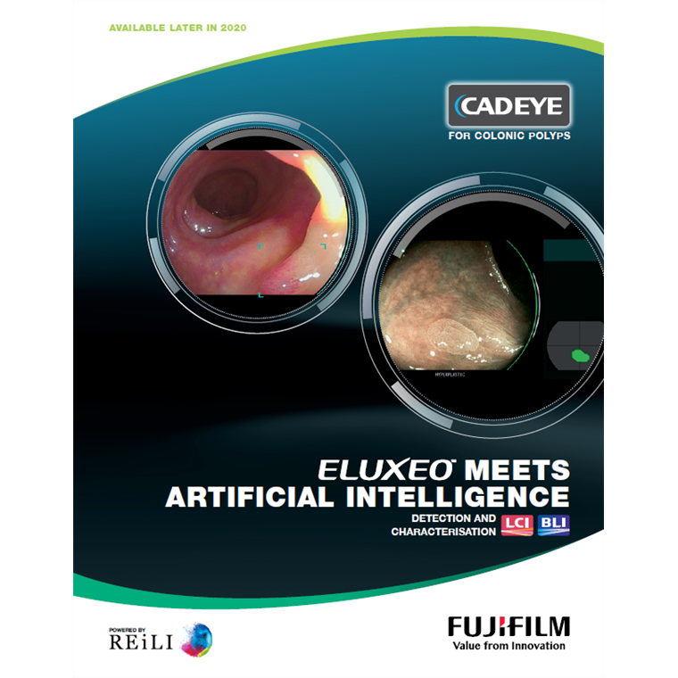
Improving the detection rate of difficult to discover lesions has been one of the major challenges within the field of endoscopy in recent years. Fujifilm has continuously worked on the development of image processing technologies such as Linked Color Imaging (LCI) and Blue Light Imaging (BLI) utilising specific wavelengths of light to support early cancer detection and characterisation. Recently Fujifilm has developed a new technology known as “CAD EYE” to support colonic polyp detection and characterisation during colonoscopy, utilising Fujifilm's medical AI technology named REiLI. CAD EYE is a customised detection and characterisation support compatible with the ELUXEO system.
Cad Eye Supports Detection & Characterisation - Customised for ELUXEO USERS
CAD EYE has been developed utilising AI deep learning technology and is compatible with Fujifilm’s ELUXEO™ endoscopy series to support endoscopic lesion detection and characterisation in the colon. CAD EYE has been trained with a powerful supercomputer located in Fujifilm’s global AI technology centre in Tokyo, utilising an immense amount of clinical images using Fujifilm endoscopy systems. As a result, CAD EYE is a customised detection and characterisation support compatible with the ELUXEO™ system.