

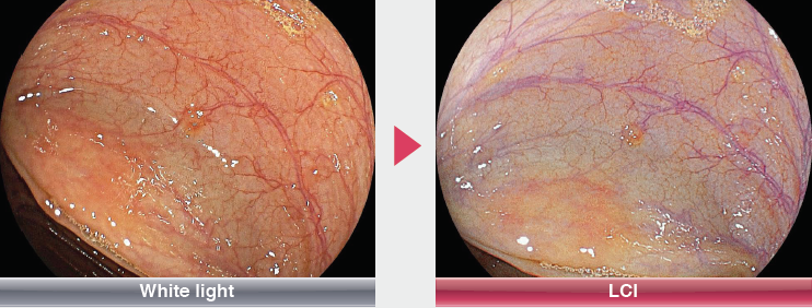
Linked Colour imaging (LCI) is a unique image-enhanced endoscopy (IEE) technology from Fujifilm Corporation that utilises a combination of blue light imaging (BLI) and white light (WL) to improve the contrast of haemoglobin in the mucosa, post signal processing then expands the colour separation, resulting in more vivid and distinct reds and whites.
The clinical benefits of LCI are described in the recent Multi-Centre, Randomised, Crossover trial extract below, which determined that overall Polyp detection with LCI increased by 24%.
Comparison of LCI and WLI for colorectal polyp detection: a multicenter, randomized, crossover trial
Editorial Supervisor: Min Min MD, PhD, Yan Liu MD, PhD, Affiliated Hospital of Academy of Military Medical Sciences, China.
New clinical evidence:
Linked Color Imaging (LCI) improves significantly adenoma detection rate (ADR) and polyp
detection rate (PDR).
Joint Research facilities & research collaborators:
Affiliated Hospital of Academy of Military Medical Sciences - Pei Deng MD, PhD
The People's Hospital of Guangxi Zhuang Autonomous Region - Wenhua Zhang MD, PhD Bing Nong MD, PhD
Shanghai Tenth People's Hospital - Xiaomin Sun MD, PhD
Background & Aim:
There is great interest in developing techniques that may improve polyp detection in the colorectal
region during colonoscopy. Several image-enhanced endoscopic (IEE) systems that enhance the
mucosal vasculature and/or architecture without the use of dye have been developed. Many clinical
studies have reported an improvement in the detection rate of colonoscopy using IEE systems.
LCI is a recently developed technology that enhances the color separation of red color to depict red
and white colors more vividly.(2) The benefits of LCI in colorectal polyp detection remain unknown.
The aim of this study was to assess the ability of LCI to increase colorectal polyp detection compared
to white-light imaging (WLI) .
Study Design:
・Study Design: A multicenter, crossover, prospective, randomized controlled trial
・Registration: May, 2016 - September, 2016
・Eligibility Criteria: Consecutive adult patients
・Equipment: Light Source LL-4450, Processor VP-4450HD / Colonoscope EC-L590WM
Procedures:
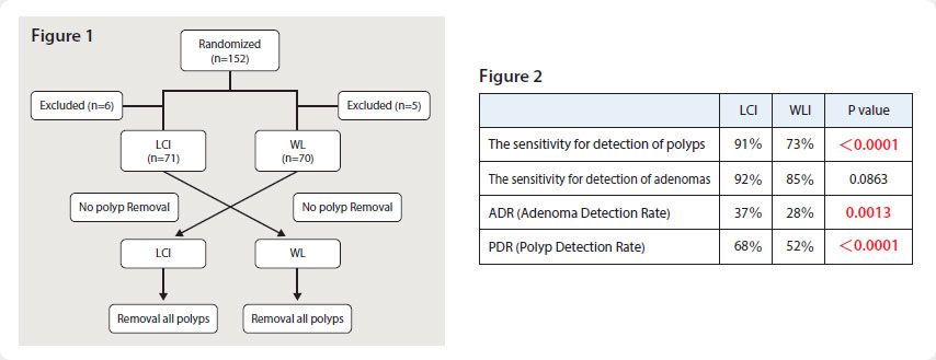
This study had a randomized, crossover design (Figure 1). Patients were randomly allocated to
1 of 2 arms: (1) LCI-WLI group: first inspection with LCI followed by a second inspection with WLI
endoscopy; or (2) WLI-LCI group. All patients underwent crossover colonoscopies with LCI and WLI
endoscopy in a randomized order. All lesions were removed during the second endoscopic procedure.
The primary outcome measure was the difference in sensitivity between LCI and WLI endoscopy for
the detection of colorectal polyps. The secondary outcome measures were the adenoma detection
rate per patient in the two groups.
Results:
A total of 152 patients were randomized, and 141 were included in the analysis.
<Sensitivity for the detection of colorectal polyps>
The overall polyp detection increased signifcantly by 24% for LCI, corresponding to a higher sensitivity of LCI than WLI (91% vs 73%, P<0.0001).
<ADR>
The ADR was signifcantly higher for LCI than WLI (ADR: LCI 37% vs WLI 28%, P=0.0013).
<PDR>
The PDR was signifcantly higher for LCI than WLI (PDR: LCI 68% vs WLI 52%, P<0.0001).
Conclusion:
LCI improves the detection of colorectal polyps and adenomas during colonoscopy.
Reference
(1): Min M1, Deng P1, Zhang W2, Sun X3, Liu Y1, Nong B2. Comparison of linked color imaging and white-light colonoscopy for detection of colorectal polyps: a multicenter, randomized, crossover trial. Gastrointest Endosc. 2017 Mar 9.
pii: S0016-5107(17)30180-3. doi: 10.1016/j.gie.2017.02.035.
(2): Okada M1, Sakamoto H1, Takezawa T1, Hayashi Y1, Sunada K1, Lefor AK2, Yamamoto H1. Laterally Spreading Tumor of the Rectum Delineated with Linked Color Imaging Technology. Clin Endosc. 2016 Mar;49(2):207-8. doi:
10.5946/ce.2015.077. Epub 2016 Feb 12.
